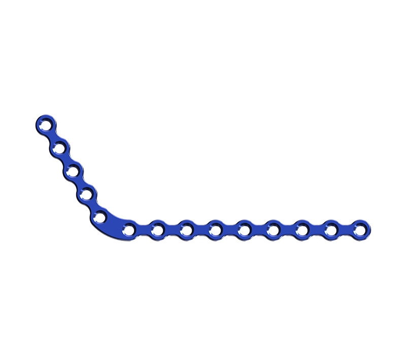
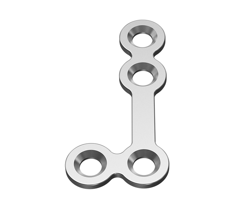
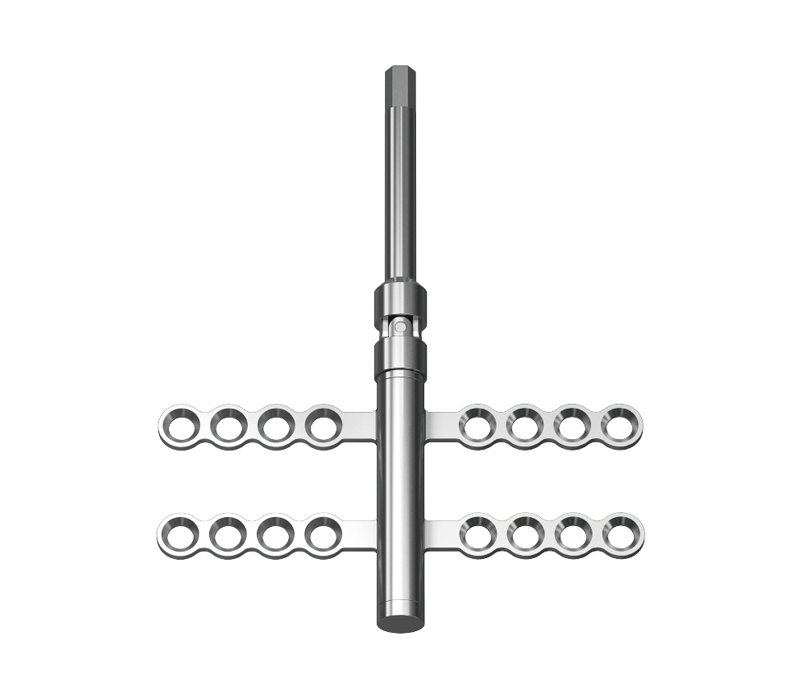
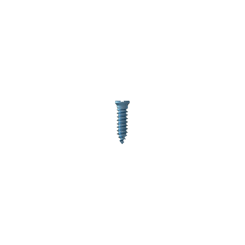
Cranial and maxillofacial fixation titanium plate
Titanium alloy screws
- Anchor nail titanium screws
- IMF Titanium Screws
- Orthodontic anchorage titanium screws
- Self-tapping titanium lag fixation screws
- Locking system of self-tapping titanium screws/emergency titanium screws
- Reconstruction of self-tapping titanium screws/ emergency titanium screws
- Self-drilling titanium screws
- Self-tapping titanium screws /Emergency titanium screws
Product center
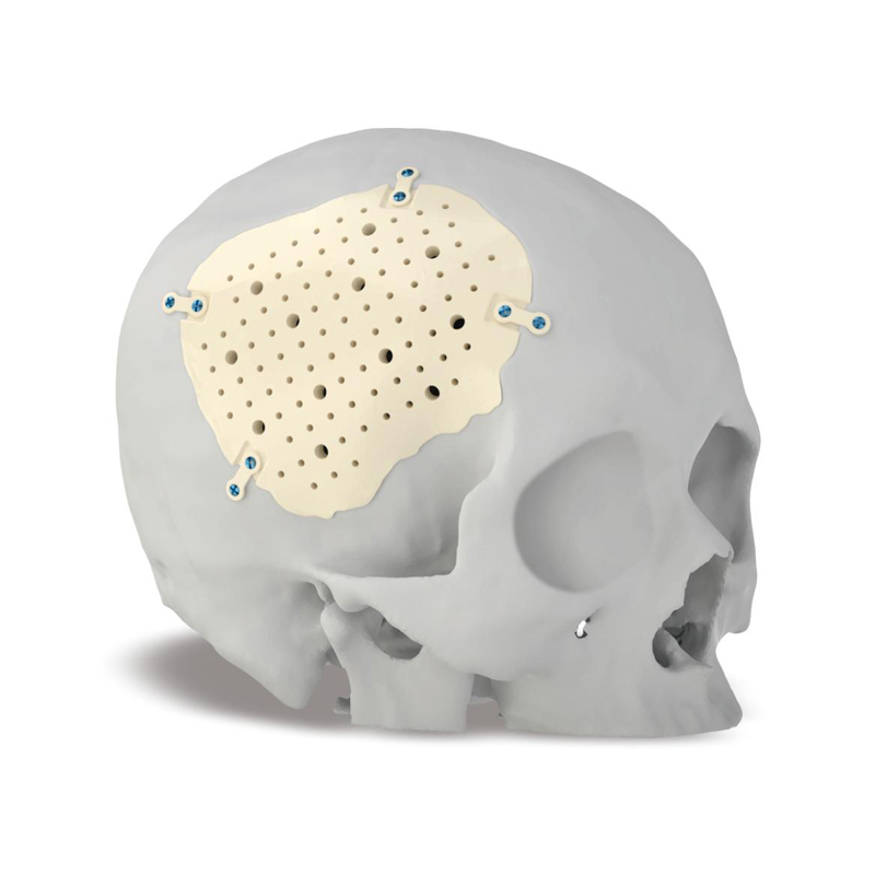
PEEK Skull Repair Body
PEEK Material Clinical Advantages
○Embedded repair and digital molding technology embed PEEK material into the bone window, achieving perfect fit with the defect area.
○Customizable bone plates of different shapes to perfectly meet anatomical reconstruction requirements.
○The mechanical properties are close to those of human bones, with a sturdy texture that is not easily subjected to stress and indentations.
○Good thermal insulation performance, reducing external temperature stimulation.

Cibei Medical is guided by market demand and serves the society as its mission. It strengthens cooperation between industry, academia and research, pays attention to refined management...
No. 8, Xiaotuan Pudong Road, East Development Zone, Guanhaiwei Town, Cixi City, Zhejiang Province
+86-0574-6366 7369
+86-0574-6366 7369
cbzj@cbzj.com.cn
Quick Links
Product center
Contact Us
您希望我们为您提供哪些服务?
Ningbo Cibei Medical Instrument Co., Ltd. all rights reserved  浙ICP备19045460号
Technical Support:HWAQ


